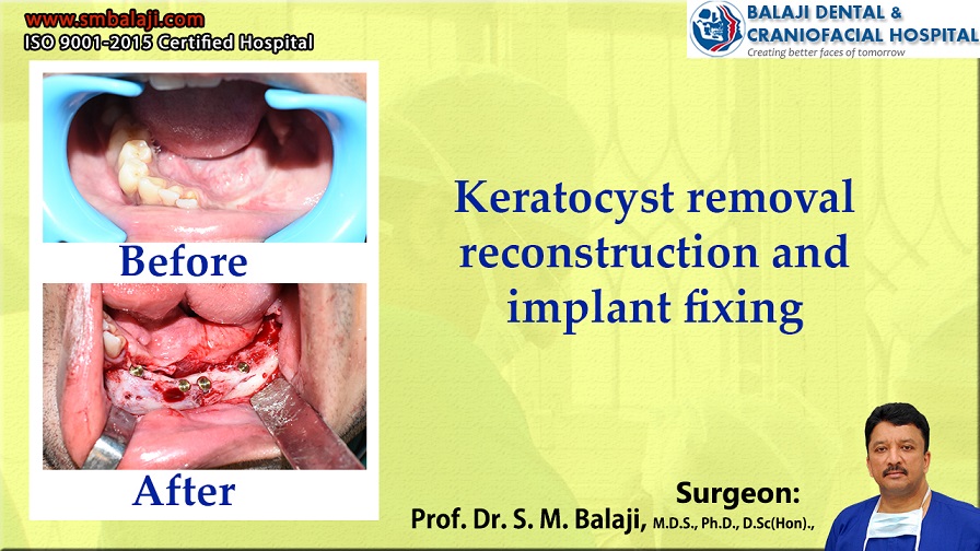
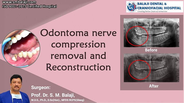
Bilateral Cleft lip and Palate – Pharyngoplasty for speech correction
Incidence of Bilateral Cleft Lip and Palate Deformity in Newborn Infants
Bilateral cleft lip and palate deformity is one of the most common congenital deformities found in infants. There is a hole in the roof of the mouth in cleft palate defect. The upper lip is split in cleft left defect. A baby with a cleft defect has feeding difficulties.
These birth defects occur at about a rate of 1 out of every 1000-2000 live births. Children with cleft lip and palate deformity have to undergo prolonged rehabilitation to reach complete normalcy. In the USA, board-certified plastic surgeons rehabilitate these children to lead normal lives. Cleft lip and palate repair enable a life of complete normalcy.
They first undergo cleft lip repair at 3-1/2 months old and cleft palate repair at 10 months old. This is followed by cleft alveolus repair at 4-1/2 years of age. Speech impediments are common in these children. They need to undergo prolonged speech therapy for correction of their speech defects.
Pharyngoplasty for Speech Correction in Children with Cleft Deformities
Most children with bilateral cleft lip and palate deformity have velopharyngeal insufficiency. This causes them to face difficulties pronouncing certain sounds. There is also a hypernasality to their voice. They thus need to undergo pharyngoplasty for speech correction once they are old enough.
Facial Plastic Surgery for Bilateral Cleft Lip and Palate Deformity
Cleft lip and palate surgery deals with functional aspects of speech and eating as well as facial esthetics. It is a very important component of facial plastic surgery. It is equally important that both the functional as well as esthetic needs of the children are met. Treatment of bilateral cleft lip and palate deformity is best performed by experienced surgeons.
Importance of Experience in Cleft Lip and Palate Surgery
The field of facial plastic surgery first underwent rapid development during the First World War. This was due to the grievous injuries suffered by soldiers on the battlefront. Many techniques of facial reconstruction first arose during this period.
Importance of Esthetic Results in Facial Plastic Surgery
The field of facial plastic surgery is not only a surgical science but also has an artistic element to it. Surgeons have to envision the end result and sculpt the facial structures accordingly. This requires an enormous amount of experience. Cleft lip and palate surgery, which often influences facial esthetics, is mastered by experienced facial plastic surgeons.
Initial Cleft Lip and Palate Surgery for the Patient
The patient is a 13-year-old female from Solapur in Maharashtra, India. She had been born with a bilateral cleft lip and palate deformity. Her parents had been counseled and provided with the right surgical schedule at the time of her birth. They had been very proactive and involved in the care of their daughter.
She had first undergone bilateral cleft lip repair when she was three months old. This was followed by cleft palate repair at 10-1/2 months old. Cleft alveolus surgery with bone grafting had been performed when she was 4-1/2 years old. Reconstructive surgery helped her achieve all her milestones normally.
This is a form of cosmetic surgery. These surgical procedures need experienced surgeons for the best results. Treatment planning commences as soon as the baby is born. The upper jaw is structurally compromised in cleft palate deformity.
Referral to Speech Therapist for Speech Correction
She stated that she had always had difficulty with the pronunciation of certain sounds. There had also been a degree of hypernasality to her speech. She had worked hard with her speech therapist to improve this, but her problems persisted.
In view of the pandemic, her school classes have been entirely online. Her teachers had raised the concern with her parents that they were having difficulty understanding her speech. Realizing the importance of this, her parents decided to get this addressed at the earliest.
Initial Consultation to our Hospital for Speech Correction
She and her parents presented to our hospital for the management of her speech problems. Various tests were performed and she was diagnosed with velopharyngeal insufficiency. Dr. SM Balaji, cleft lip and palate surgeon, explained the treatment planning to them. They were in complete agreement with the proposed treatment plan and consented to surgery.
Pharyngoplasty for Speech Correction performed for the Patient
It was decided to perform a pharyngoplasty for speech correction. The procedure was explained to them. It was explained to them that she would need further speech therapy for the complete rehabilitation of her speech problems.
Successful Resolution of Hypernasality of her Speech
An Orticochea dynamic sphincter pharyngoplasty was performed for her. This surgery is performed at the back of the palate. There was a creation of a small central port in her soft palate to facilitate normal nasal breathing.
A suction test was performed at the end of the surgery. This demonstrated completely normal action of the soft palate as opposed to the velopharyngeal insufficiency before surgery.
There was a remarkable improvement in the quality of her speech following surgery. As explained previously, her rehabilitation process would continue further with her speech therapist. This would result in complete resolution of the problems caused by her bilateral cleft lip and palate deformity
Total Satisfaction with the Treatment Process at our Hospital
She and her parents expressed their complete satisfaction with the level of care at our hospital. They thanked the members of the surgical team before her final discharge from the hospital.