Warning: Constant WP_CACHE already defined in /home/smbalnoi/backup.drsmbalaji.com/wp-config.php on line 86 Surgery – Page 7 – Balaji Dental and Craniofacial Hospital, Chennai, India
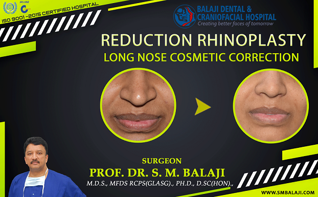
Young lady who has long desired to undergo cosmetic nose correction for long nose
The patient is a 23-year-old female from Nagpur in Maharashtra, India. She has always felt that her nose was not in harmony with her cheek bone structure. Feeling that it did not suit her facial features, she had long desired long nose cosmetic reduction surgery.
Her parents had said that she needed to finish her studies before she underwent this procedure. Having finished her studies, they decided that this was the ideal time to do this.
They had approached a leading plastic surgeon in her hometown for this. He undertook various facial analyses to study her nose and its relationship to her face. Realizing that this needed to be performed by a rhinoplasty specialty surgeon, he had referred them to our hospital.
Our hospital is a leading facial cosmetic surgery hospital in India. We perform both cosmetic nose surgery as well as nasal deformity surgery. Cleft rhinoplasty correction is a superspecialty offering in our hospital. Cleft lip nasal deformity rehabilitation has restored the lives of many patients. We have two state of the art operating theaters for performing all surgeries.
Many successful celebrities from fields as diverse as media, films and sports have undergone corrective rhinoplasty at our hospital. The patient and her family immediately got in touch with our hospital manager. She said that she wanted a cosmetic nose job. They were asked to report to our hospital for an initial consultation.
Initial presentation at our hospital for definitive correction of her long nose deformity
Dr SM Balaji, cosmetic rhinoplasty specialist, examined the patient and obtained a detailed history. She expressed her displeasure at the length of her nose. Another issue that bothered her was the breadth of her nostrils, which she felt were excessively flared. She desired to undergo cosmetic correction of these two issues. A closed rhinoplasty would be the ideal approach to avoid scar formation.
He then ordered detailed facial measurements along with nasal dimensions for the patient. Various cephalometric and other parameters were then utilized to determine the best nasal form for the patient’s face. The patient was educated about cosmetic surgery and what to expect from it. Time was spent listening to the patient to understand her expectations better.
A detailed treatment plan was then formulated to address the patient’s issues. This would involve reduction of the nasal cartilages to reduce the length followed by Weir excision for alar reduction. The procedure was explained to the patient and her parents who consented to the surgery.
Successful reduction of the length of nose to a perfectly esthetic form
The patient was prepped and draped for surgery. Careful measurements were made pertaining to all the surgical landmarks. Under general anesthesia, intranasal incisions were made and the lower lateral cartilages were reduced.
This was then followed by reduction of the upper caudal part of the nasal septum. A T-shaped strip of cartilage was then removed through an intranasal transcartilaginous incision to reduce the length of the nose. This was followed by stripping of the perichondrium. All incisions were then closed with sutures.
Attention was next turned towards correction of the broad alar base. Measurements were made and this was then corrected through a Weir excision procedure. Incisions were again closed with sutures. This resulted in a nasal form that was in perfect harmony with the patient’s facial features.
Total patient satisfaction with the results of the cosmetic nasal correction
The patient who works in the media was ecstatic over the results of the surgery. She expressed that she now has the nose that she had always desired. Her parents were also very happy with the results of the surgery.
She said that she would now be able to concentrate on her profession with a greater level of self confidence. The patient stated that being in the media required being confident about one’s appearance. They expressed their heartfelt gratitude before final discharge from the hospital.
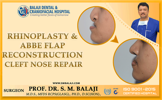
Past history of surgical correction of congenital cleft lip, palate and alveolus defect
The patient is a 21-year-old female from Ongole in Andhra Pradesh, India. She was born with a bilateral cleft lip, palate and alveolus defect. There was an oronasal communication through the roof of the mouth. Her parents were extensively counseled at the time of birth. They were then referred to our hospital for surgical management.
Her parents followed instructions meticulously and she underwent bilateral cleft lip repair at 3 months of age. The initial surgery was followed at 8 months by cleft palate repair. She also underwent premaxillary setback with bone grafts to the cleft alveolus at 7 years of age. A sphincter pharyngoplasty was performed at 3-1/2 years to address her speech problems.
Orthodontic intervention at our hospital for correction of malaligned teeth
She has been under the care of an orthodontist since the age of 9 at our hospital. Rapid palatal expansion had been done for the patient to correct her posterior crossbite. She also underwent fixed orthodontic treatment for correction of her malaligned teeth. The patient also had congenitally missing maxillary teeth due to her cleft alveolus deformity.
With the passage of time as the patient grew up, her nasal deformity however became more pronounced. Her columella was collapsed to the point that it appeared to be absent.
This caused her nose to be extremely flattened and blunt. Her prolabium was also extremely stunted. She would require a nose job. No nasal tip graft would be required in this case. It was therefore decided to surgically correct her cosmetic nasal defects.
Plastic surgeons also perform this surgery in the Western countries. Head neck specialists are often cosmetic as well as maxillofacial surgeons. They need to be board certified to perform these surgeries. Reshaping of bone and cartilage is often required for many facial plastic surgeries.
Presentation for nasal cosmetic surgery to be performed at our hospital
Dr SM Balaji, cleft cosmetic surgeon, examined the patient. She had an extremely shortened columella along with a stunted prolabium. Meticulous treatment planning was done for the patient. It was decided that both the issues needed to be addressed.
The congenitally missing maxillary teeth would be addressed later through the placement of dental implants. Her columella would be reconstructed utilizing a flap taken from the prolabium. This would be followed by an Abbe flap reconstruction of the ensuing upper lip defect.
The patient and her parents were in complete agreement with the surgical plan and consented to surgery.
Surgical repair of nasal and lip defect using Abbe flap reconstruction
Under general anesthesia, incisions were made in the region of the stunted prolabium. This was followed by dissection of the columella. The medial and lateral crus of the nasal septum were then identified. These were then brought together to form the alar dome.
This would help in creating a more prominent and sharp alar dome. The prolabium was then utilized to reconstruct the columella and lengthen it.
This resulted in a defect in the region of the prolabium. A full thickness Abbe flap was raised from the lower lip. This flap was then sutured to the defect in the region of the prolabium. Care was taken to ensure that the inferior labial artery remained patent throughout the procedure.
The patient would not be able to open her mouth for two weeks. During this time, the flap would be deriving its blood supply from the inferior labial artery of the lower lip. Once vascularity to the flap had been established from upper lip blood vessels, it would be separated from the lower lip.
Surgical separation of the lower lip Abbe flap after establishment of vascularity
The flap was separated from the lower lip and demonstrated good blood supply from the upper lip. She now had a prominent nose with a long columella as well as a lengthened prolabium. The patient and her parents were very happy with the results of the surgery. They thanked the surgical team and explained that this would transform her life before final discharge.
The patient involved in a head-on car crash while on a work trip
The patient is a 26-year-old businessman from Pune in Maharashtra, India. About a year ago, he had driven down to a nearby city for work. While there, he was involved in a head on collision with another automobile. His head had impacted directly on the steering wheel resulting in extensive lower facial injury. He had not been wearing his seatbelt at the time of the accident.
He suffered a fractured lower jaw along with fractured teeth in both jaws. An ambulance had been summoned and he was immediately shifted to a nearby hospital. Imaging studies obtained at the hospital revealed bilateral body of the mandible fractures near the premolar region. He had also suffered fractures to maxillary and mandibular anterior teeth.
There were no injuries to his eye sockets and bone fractures to other facial structures. This type of injury is very common to unrestrained drivers. They can be life threatening and surgeons with special training are required to address these complex injuries.
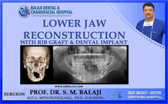
Complications of malunion and infection with resultant loss of alveolar bone
An emergency surgery had been performed and his bilateral mandibular fractures had been stabilized with titanium plates. His fractured anterior teeth were also extracted.
Within a month after surgery, it became evident that the surgery had been poorly performed. Bilateral fracture fixation sites developed an infection. This resulted in extensive resorption of the anterior alveolar bone. The height of the alveolus was greatly reduced.
Referral to our hospital for management of complications of previous surgery
The patient presented at a local hospital for management of his problem. Imaging studies were obtained. The patient was informed that he had suffered from extensive alveolar bone loss secondary to the infection.
There was malunion of the fracture repair with infected plates. Postsurgical suturing had been performed poorly with resultant abnormal frenal attachments.
The patient and his family were counseled extensively regarding this. It was explained to them that his problem needed to be addressed at a specialty facial trauma care hospital. They were then referred to our hospital for treatment.
Anxiety of patient and his family allayed during initial consultation in our hospital
Dr SM Balaji, facial trauma care surgeon, examined the patient and ordered imaging studies including a 3D CT scan. There was malunion of the fracture along with reduced alveolar height. This had been caused due to the infection at the fracture site. There was also extensive loss of anterior teeth in both the maxilla and the mandible.
Treatment planning explained in detail to the patient and his family
It was explained that the patient’s infected titanium plates needed to be removed. This would be followed by reconstruction of the alveolar bone using bone grafts harvested from the patient. Nobel Biocare dental implants would then be placed for rehabilitation of the missing upper anterior teeth.
A period of six months would be allowed for bony consolidation of the graft to the alveolar bone. This would be followed by the placement of dental implants for rehabilitation of the missing lower anterior teeth.
Ceramic crowns would be placed on the dental implants following complete osseointegration of the implants to the surrounding alveolar bone. The patient and his parents were in complete agreement with the treatment plan and consented to surgery.
Successful completion of jaw reconstructive surgery for the patient
Under general anesthesia, an inframammary incision was made and a rib graft was harvested. A Valsalva maneuver was then performed to ensure that there was no perforation into the thoracic cavity.
Attention was turned to the maxilla where dental implants were placed in the anterior region. This was followed by a mandibular vestibular incision with the removal of the infected plates. The rib graft was then crafted to fit perfectly into the anterior alveolar defect. This was then fixed in place with screws and flaps closed with sutures.
Patient expresses understanding of postoperative instructions
The patient and his parents expressed their satisfaction with the treatment planning and the surgical results. They stated that they will return in six months for the complete rehabilitation of his oral structures.
Patient develops pain and swelling in the posterior region of his right mandible
The patient is a 36-year-old man from Satna in Madhya Pradesh, India. He had developed pain and swelling in the molar region of his right lower jaw about six months ago. This had gradually increased in size with slight mobility of the first and second molars.
History of small cell lung cancer secondary to an extensive history of smoking
The patient is a cancer survivor. He had been diagnosed with carcinoma of the lung at the age of 32 years. Secondary to the diagnosis, he had immediately stopped smoking. Starting from the time of diagnosis, he had received chemotherapy and radiotherapy for a period of two years at a nearby city.
His cancer has been in remission since then and he is regularly followed by his oncologist. Of all the types of lung cancer, small cell cancer responds best to multidrug chemotherapy regimen.
Alarmed at the development of pain and swelling due to his history of cancer, he had rushed to his oncologist for advice. His oncologist had examined him and obtained an OPG. Realizing that the patient had a cystic lesion, the oncologist made widespread enquiries regarding the best hospital for addressing this new development.
Having received multiple recommendations about our hospital, he looked up our hospital on the Internet and spoke with Dr SM Balaji, Director and Consultant Oral and Maxillofacial Surgeon of our hospital. The patient had then been referred to our hospital for management of his cystic lesion. Our hospital is also a specialty center for orthognathic surgery.
Patient presents at our hospital for management of his right jaw swelling
Dr SM Balaji, dentigerous cyst surgeon, examined the patient and ordered imaging studies including a 3D CT scan. This revealed the presence of an impacted third molar in relation to the cystic lesion. There was extension of the cystic lesions up to the roots of the first and second molars. A biopsy confirmed the diagnosis of a medium to large dentigerous cyst.
In view of the patient’s history of treatment for carcinoma of the lung, it was decided that treatment would only address the cystic lesion. This would involve enucleation of the cystic lesion with fixation of a mandibular reconstruction plate. It was decided to utilize an intraoral approach to perform this surgery to enable easy healing of tissues.
Patients with dentigerous cyst usually undergo complete rehabilitation at our hospital. Treatment would include placement of rib grafts obtained from the patient followed by placement of dental implants. This would result in full restoration of function for the patient.
Successful surgical enucleation of dentigerous cyst with extraction of involved molars
Under general anesthesia, a vestibular incision was made in the right molar region of the mandible. Dissection was carried down to the region of the dentigerous cyst. There was slight perforation of the buccal cortical bone. The impacted third molar was removed followed by enucleation of the cystic cavity.
Care was taken to ensure safety of the inferior alveolar and mental nerve throughout surgery. A Titanium mandibular reconstruction plate was then used to reconstruct the mandible. The incision was then closed with sutures and the patient recovered uneventfully from the surgery.
Nerve function testing was done and was found to be intact. The patient and his family were fully satisfied with the results of the surgery. They expressed their thankfulness to the surgical team before final discharge from the hospital.
Patient born with left-sided cleft lip, palate and alveolus deformity
The patient is a 19-year-old male from Thanjavur in Tamil Nadu, India. The product of a consanguineous marriage, he was born with a unilateral cleft lip, palate and alveolus deformity. His parents were referred to a dental surgeon in the hospital who explained the treatment protocol to them. They had then been referred to our hospital for management.
Upon arrival at our hospital, an examination was performed followed by a comprehensive treatment plan. He had subsequently undergone cleft lip surgery at 3 months and cleft palate surgery at 8 months. His cleft alveolus deformity had been addressed with a bone graft at 7 years. He had been under the care of an orthodontist from the age of 10 onwards.
Development of maxillary retrusion with a resultant sunken face appearance
As the patient grew up, he noticed that his maxilla was getting retruded. This was caused by a hypoplastic maxilla, which resulted in a sunken face deformity to his face. There was also the gradual development of a skeletal anterior crossbite. This had resulted in speech and eating difficulties.
There was also a degree of compromise to his facial esthetics. He felt that it made him look abnormal. This was causing him to be socially withdrawn and depressed.
The patient discussed this with his parents and they decided to return to our hospital to address this. Our hospital is a premier center for facial cosmetic surgery in India. Our hospital has always been at the forefront of advancements in orthognathic surgery in India.
Scores of patients have benefitted from the facial esthetic surgery performed in our hospital. Reconstructive facial plastic surgery is also performed in our hospital for the facial region. Sunken cheeks are usually filled out when orthognathic surgery is performed for retruded jaws.
Patient and his parents present for correction of his sunken maxillary defect
Dr SM Balaji, Le Fort I surgery specialist, examined the patient and obtained imaging studies including a 3D CT scan. The patient had a skeletal anterior crossbite. It was explained to the patient that he needed to undergo maxillary advancement surgery to correct this problem. The patient and his parents expressed understanding of the treatment plan and consented to undergo surgery.
Successful surgical correction of his midfacial maxillary deformity
Under general anesthesia, a vestibular incision was made in the maxillary sulcus. A Le Fort I osteotomy was then performed and the maxillary bone disjointed. The maxillary bone was then advanced forward and occlusion was checked.
This was followed by fixation of the maxillary bone with titanium plates and screws. The incision was then closed with sutures. Anesthesia was successfully reversed and the patient was taken to the recovery room in stable condition.
Complete patient satisfaction with the results of the surgery
There was an immediate improvement in the patient’s facial esthetics following surgery. He now had a pleasing facial profile following forward advancement of his maxilla. The patient and his parents said that he would now develop more self confidence. They expressed their thankfulness before final discharge from the hospital.
Patient with long-standing poorly controlled diabetes mellitus
The patient is a 48-year-old male from Sambalpur in Odisha, India. He has a very strong family history of diabetes on both the paternal and maternal sides of his family. Diagnosis of early onset type 2 diabetes mellitus had been at the age of 28. He had however never shown any inclination to control it. His blood sugar levels regularly went off the charts and he has required innumerable hospitalizations in the past.
Typically, type 1 diabetes mellitus is diagnosed earlier in life. It is usually much more severe than late adult onset diabetes mellitus.
Members of his family had warned him about the consequences of his lifestyle choices, but he had ignored them. Doctors had warned him about the ophthalmological, neurological and renal consequences, but he disregarded medical advice. Secondary to his diabetes and long standing poor oral hygiene, he developed destructive periodontal disease in his early thirties.
Ignoring the warnings of his dentist, he had lost most of his mandibular teeth within a few years. This very early edentulous state resulted in early resorption of his mandibular bone. His maxillary teeth were in no better shape and had severe gingival recession.
Patient decides to make major lifestyle changes for enhanced quality of life
It has only been over the last two years that the patient has decided to lead a disciplined life. He started exercising regularly and strictly monitoring his dietary intake. His blood sugar levels have been normal for over a year now. There has been a tremendous improvement in his energy levels and his overall zest for life.
Poor oral health leading to early loss of majority of teeth
The patient however had already paid a heavy price with regards to his oral health. He had already lost most of his mandibular teeth and all his maxillary teeth were mobile. There was also a tremendous amount of alveolar resorption of the mandibular bone, which gave him a prematurely aged look.
Feeling depressed about this, he had approached a local dental surgeon for dental implants. Imaging studies had been obtained at the consultation. It was then that the dentist realized the complicated presentation due to the degree of alveolar resorption of the mandible. He explained the complexities to the patient and referred the patient to our hospital for treatment.
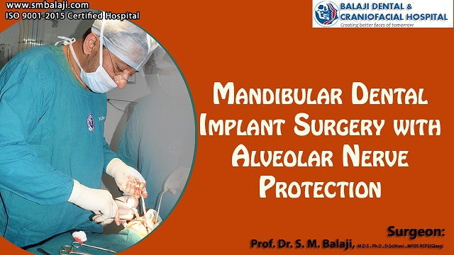
Dental implant surgery services available at our hospital
Our hospital is a leading specialty dental implant hospital in India. We specialize in total oral rehabilitation with the use of a variety of dental implants. Graftless zygoma implant surgery is utilized for patients who do not wish to undergo surgery under general anesthesia. Zygomatic implants enable rehabilitation of maxillary arch without grafts.
Over a 1000 patients have been successfully rehabilitated with restoration of a beautiful smile. Our hospital was one of the first to offer implant dentistry in India. Tooth replacement is most convenient for the patient with implants, particularly for full mouth rehabilitation. Implantology is also a subdivision of maxillofacial surgery. Conventional implants need bone grafts in case of severely resorbed arches.
Complications of his presentation explained during treatment planning
Dr SM Balaji, dental implant specialist, examined the patient and obtained imaging studies including a 3D CT scan. He explained to the patient that the lower arch would be addressed first. Imaging revealed that there was significant resorption of the mandibular alveolar ridge. This would result in upward migration of the mental nerve towards the ridge.
It was explained to the patient that implant placement distal to the mental nerve could be easily accomplished. The patient was made to understand that proximal implants could be placed only after careful mapping of the mental nerve. This would ensure that there was no nerve damage during placement of the implants.
The patient and his family expressed understanding of the treatment plan and consent to undergo surgery.
Successful surgical placement of implants without resultant nerve damage
Under general anesthesia, a supracrestal incision was made and a flap was raised. The alveolar crestal bone was exposed and mental nerve identified. Six Nobel Biocare dental implants were carefully affixed for rehabilitation of the mandibular dentition. The flap was then closed with sutures followed by removal of the mobile maxillary teeth.
Postoperatively, there were no signs of numbness or symptoms of damage to the nerve. Instructions were given to the patient to return in six months after completion of osseointegration of his mandibular implants. It was explained that maxillary rehabilitation will be undertaken at a later date.
The patient and his family expressed understanding of the instructions and thanked the surgical team before discharge from the hospital. The importance of keeping his blood sugar levels under check were reiterated to the patient. He stated that he has now made drastic lifestyle changes. This would enable him to enjoy greater health in the days to come.