Salivary Gland
Salivary glands are glands found in and around the mouth and throat. They secrete saliva into the mouth through tubes called salivary ducts. There are 3 major salivary glands- the parotid, submandibular and sublingual glands.
Besides these, there are 600-1000 tiny glands called minor salivary glands located in the lips, inner cheek area (buccal mucosa), and linings of the mouth and throat. Salivary glands produce saliva that moistens the mouth, acts as a lubricant for speech, helps in swallowing, initiates digestion, has antibacterial properties and protects teeth from decay.

Diagrammatic representation of the location of the major salivary glands
Salivary gland diseases
Disorders of the salivary glands include
1. Hyperplasia or swelling of the salivary glands:
Rarely, the minor salivary glands may be swollen. They are seen as mild, painless masses usually on the roof of the mouth and soft palate. A biopsy is needed to be done to rule out other diseases. Once diagnosed, these swellings may subside on their own.
2. Salivary stones or calculi in the gland causing obstruction to the saliva flow
Sialoliths or salivary gland stones may be formed in the salivary glands or ducts. These cause obstruction to the flow of saliva. Patients complain of periodic painful swelling, typically when eating. The salivary glands swell then gradually subside after eating. If not treated early, infections or abscesses may occur.
Sometimes the salivary ducts may have small constrictions, which decrease salivary flow, leading to infection (sialadenitis) and obstructive symptoms.
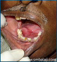
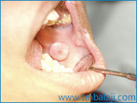
A bulge on the right side of the floor of the mouth indicating the presence of submandibular gland calculus

A bulge on the right side of the floor of the mouth indicating the presence of submandibular gland calculus
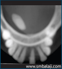
Occlusal radiograph of the same person showing an oblong radiopacity of the calculus in the right side of the mandible

Excised calculus (salivary gland stone)
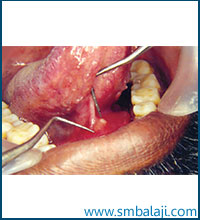
Submandibular salivary gland calculus present at the entrance of the duct

The excised calculus
3. Salivary gland infections
Infections in the salivary gland may be viral, most commonly mumps, or bacterial. Most bacterial infections occur due to obstruction of salivary flow. Secondary infections of salivary glands from nearby lymph nodes may also occur. The affected gland gets swollen and painful accompanied with fever and difficulty in opening the mouth. Treatment includes antibiotic therapy and surgical drainage of the infection in case of abscess.
4. Trauma to the duct causing its rupture
Sometimes due to injury, the salivary gland duct may rupture leading to spillage of mucin into the surrounding soft tissues. This leads to the formation of mucocele or small dome-shaped swellings. These are very commonly seen on the lower lip or cheek. When they occur on the floor of the mouth, they are called ranula (retention cyst).

Mucocele in the lower lip
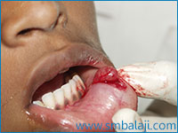
Mucocele in the lower lip excised
Mucocele in the region of the left cheek

Ranula (retention cyst)
5. Under-functioning of the salivary gland causing dry mouth or xerostomia
Dry mouth may be due to various causes like improper development or functioning of the salivary glands, excess water loss from the body, as a side effect of some medications or radiation therapy to the head and neck, diseases like Sjögren’s syndrome, Diabetes or dry mouth may also occur due to local factors like smoking and mouth breathing. To cure the mouth dryness, first the underlying cause must be treated. Salivary substitutes may be used, or sugarless candy or gum can help keep the mouth moist. Good oral hygiene should be maintained to prevent mouth infections.
6. Salivary gland tumors
Benign or malignant tumors of the salivary glands may occur as growth or enlargements in the roof or floor of the mouth, cheek or lip. One or more glands may be involved. Any pain or loss of function of the affected region should be immediately investigated.
7. Autoimmune diseases like Sjögren’s syndrome
In some autoimmune diseases like HIV and Sjögren’s syndrome, the salivary glands may be inflamed. Along with dry mouth (xerostomia) there may also be painful and burning sensation and dryness of the eyes. Secretory glands of the nose and throat may also be involved. Treatment is mostly supportive. Tear and saliva substitutes may be used to prevent dryness. Increased risk of tooth decay and fungal infections should be managed with preventive care.
Tests and investigations for salivary gland diseases
We advocate the latest advanced imaging techniques to diagnose and treat salivary gland diseases. The various imaging techniques include Sialography (specialized dye is injected in the salivary gland and an X-ray of the gland is taken), Computed Tomography (CT Scans), Ultrasonography, Magnetic Resonance Imaging (MRI), radioisotope scanning.

Sialography performed in submandibular gland
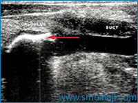
Ultrasound showing the presence of calculus in the salivary gland ductd
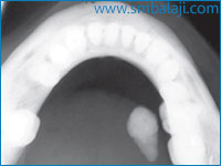
X-ray of the mandible (lower jaw) showing a mass in the floor of the mouth on the right side denoting a submandibular gland stone
If an obstruction of the major salivary glands (salivary gland stone) is suspected, dental x-rays may show where the calcified stones are located. Sometimes, a biopsy is needed to be done of the salivary gland swelling.
Treatment of salivary gland diseases
Treatment of salivary diseases includes medical and surgical modalities. Type of treatment depends upon the nature of the disease. If it is due to underlying medical conditions, then that must be treated first.
Some salivary gland lesions require to be removed surgically. Salivary gland stones or sialoliths have to be surgically excised. If the stones are located within the duct, surgical approach through the mouth may be planned. If the stones are within the gland and if there are severe changes in the duct structure, then the gland may be surgically removed extraorally.
In case of abscess, surgical drainage of the pus with antibiotic therapy may be required.

Incision placed for removal of submandibular gland

Submandibular gland removed

The excised gland

The wound sutured

Surgical removal of the parotid gland
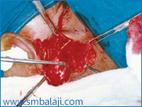
Submandibular gland removed
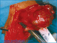
Surgical removal of parotid gland
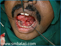
Salivary gland sialolith (salivary gland stone)
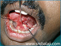
During removal of the sialolith

During removal of the sialolith
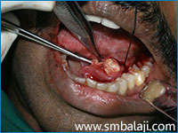
The sialolith being removed
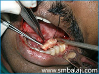
The stone surgically removed

Excised stone
If an abnormal growth or mass has developed within the salivary gland, removal of the mass may be recommended. During surgery, great care must be taken to avoid damage to the facial nerve. This is an extremely vital nerve of the face that is responsible for movements of the facial muscles including the mouth and eye.


